The work presented here supports core, the HCV capsid protein as a novel target for anti-HCV drug development. We show that an inhibitor of capsid protein dimerization can specifically and directly bind to core and core-based complexes with other HCV proteins. This binding possibly results in disruption of assembly or in disassembly of the viral particle, leading to reduction of infective HCV particles. One added advantage of HCV core over the other currently identified targets is its remarkable conservation among all six genotypes,  especially in the previously described “homotypic” region of dimerization. Inhibitors optimized on the basis of analogues described here have been found to be equally active on core proteins of genotype 1a or 1b and to inhibit virus production of a HCV 2a strain at nanomolar concentration. Despite several attempts, no resistance mutant were so far found to emerge rapidly in HCV 2a-infected cells grown in the presence of increasing concentrations of core inhibitors. Transketolase is a homodimeric enzyme that catalyses the reversible transfer of two carbons from a ketose donor substrate to an aldose acceptor substrate. Transketolase is the most active enzyme involved into the non-oxidative branch of the pentose phosphate pathway, in charge of generating the ribose molecules necessary for nucleic acid synthesis. Together with the finding that this pathway is highly expressed in the cancer cell, this enzyme provides an excellent target for novel chemotherapeutic agents. Additionally, several crystal structures of this enzyme are available and notably, the human variant of transketolase was recently reported as well allowing the rational structure-based design of human inhibitors. The active centre of transketolase contains a thiamine pyrophosphate cofactor, coordinated to a divalent metal ion, whose binding site has been used for the development of enzyme inhibitors. The most representative inhibitors that mimetize the interactions of thiamine pyrophosphate are oxythiamine and thiamine thiazolone GDC-0199 1257044-40-8 diphosphate. Unfortunately, these compounds lack selectivity as thiamine pyrophosphate is a common cofactor found in multiple enzymes, such as pyruvate dehydrogenase. More recently, several thiamine antagonists were designed with the aim of obtaining more selective inhibitors with Bortezomib improved physical properties. Nonetheless, it is interesting to find additional binding sites allowing drug discovery, not based on the active centre of transketolase but on critical allosteric points of the enzyme. Here, we utilize the homology model of human transketolase recently reported by our group to analyze the hot spot residues of the homodimeric interface and perform a pharmacophore-based virtual screening. This strategy yielded a novel family of compounds, containing the phenyl urea group, as new transketolase inhibitors not based on antagonizing thiamine pyrophosphate. The activity of these compounds, confirmed in transketolase cell extract and in two cancer cell lines, suggests that the phenyl urea scaffold could be used as novel starting point to generate new promising chemotherapeutic agents by targeting human transketolase. During the course of this research, the crystal structure of human transketolase was made public allowing its comparison with our previously reported homology model that was used in the virtual screening protocol. Nonetheless, this sequence is solvent exposed not participating in dimer stabilization nor catalytical activity. It is worth mentioning that the proposed pharmacophore used in this study can be also extracted, with minor distances differences, from the crystal structure of human transketolase.
Author: neuroscience research
The relatively smaller effect of radiation with rapamycin in contrast with the observed synergistic effect of radiation with sunitinib
The antiangiogenic effects were attributed to BAY-60-7550 decreased production of VEGF and resistance of endothelial cells to VEGF stimulation. Further studies used the rapalog RAD001 and compared its effects with known antiangiogenic agents. The results showed that RAD001 was found to be associated with decreasing the tumor vessel density and the maturity of the tumor vessels, whereas the antiangiogenic  drug vatalanib was found to impact only the microvascular density but not the vessel maturity consistent with this class of drugs which impact the VEGF/ VEGFR complex. On the other hand, Zhang et al. reported that the aSMA level, a marker of mature pericytes, increased in Tubulin Acetylation Inducer rapamycin treated tumor compared with non-treated tumor. Other studies have shown that radiation induces activation of mTOR pathways in the tumor endothelial cells making them more sensitive to response with rapamycin. However, a more recent study using a retro-inhibition approach found that HNSCC cells and not the tumor microenvironment as the target for rapamycin activity and that the anti-angiogenic effect is a likely downstream consequence of mTOR inhibition in cancer cells. Imaging of properties intrinsic to tumor physiology such as tumor pO2 and tumor microvessel density made it possible to sequentially follow rapamycin induced changes during the course of treatment non-invasively and sequentially during the treatment course. The key finding in the present study pertaining to the rapamycin effect on tumor physiology is that the tumor microvessel density, when monitored longitudinally showed a significant decrease whereas a transient increase in tumor pO2 was found followed by onset of hypoxia. It should be noted that the USPIO-based blood volume assessment may overestimate the values in tumors because of their leakiness compared to normal tissues. The observations from histological experiments were in agreement with the imaging observations. The rapamycin-induced decrease in CD31 staining was found to be in agreement with imaging experiments where a loss in microvessel density was found. However, there was a small but non-significant decrease in staining of aSMA which reflects the retention of the integrity of the pericyte coverage of the tumor vasculature after rapamycin administration. These results indicate that rapamycin treatment pruned immature blood vessels rather than mature blood vessels. It is expected that these changes in tumor microvasculature can cause improvement of blood flow, a phenomenon known as vascular normalization. The transient increase in the pO2 by rapamycin treatment can be attributed to the increased blood flow in the tumor, which was demonstrated by a 40% increase in tumor initial uptake of Gd-DTPA 2 days after rapamycin treatment in the DCE-MRI study. The identification of transient improvements in tumor oxygenation 2 days after rapamycin treatment provides an opportunity for chemoradiation modalities where radiation therapy can be timed to take advantage of increases in tumor pO2 to elicit improved response. The results in the present study show enhancement in tumor radioresponse by rapamycin treatment. This data suggests that the transiently increased level of median tumor pO2 in rapamycin treated mice compared to the day matched control group may be responsible for the observed effect of radioresponse with combination treatment.
Drug development around PTP1B has provided a proof-ofconcept for investigations focused on additional PTP targets
Several studies have uncovered physiologically important and disease relevant functions for the classic receptor type PTP, PTPs, which underscore its potential as a biological target. PTPs is highly AZ 960 expressed in neuronal tissue where it regulates axon guidance and neurite outgrowth. Furthermore, it was recently reported that loss of PTPs facilitates nerve regeneration following spinal cord injury, owing to the interaction of its ectodomain with chondroitin sulfate proteoglycans. In addition to its neural function, PTPs has been implicated in chemoresistance of cancer cells. First, we discovered that RNAi-mediated knockdown of PTPs in cultured cancer cells confers resistance to several chemotherapeutics. Additionally, we have discovered that loss of PTPs hyperactivates autophagy, a cellular recycling program that may contribute to chemoresistance of cancer cells. Taken together, it is apparent that modulation of PTPs may have therapeutic potential in a range of contexts. Notably, inhibition of PTPs could potentially provide benefit following SCI through enhanced neural regeneration. In addition, it is possible that PTPs inhibition may yield therapeutic value in diseases in which increasing autophagy represents a promising treatment strategy. Furthermore, a small Nutlin-3 moa molecule would provide value as a molecular probe or tool compound to interrogate the cellular functions and disease implications of PTPs. Several approaches exist for the identification of small molecule inhibitors of phosphatases. While high-throughput screening of compounds in vitro has been successfully utilized to discover inhibitors of LAR, PTP1B, SHP2, CD45, and others, the technical and physical investment is considerable as is the potential for experimental artifacts leading to false negatives and positives. Alternatively, a primary screen for inhibitor scaffolds can be guided by in silico virtual screening. This method involves high-throughput computational docking of small molecules into the crystal structure of a phosphatase active site and selecting the molecules which bind favorably, akin to a natural substrate. Following the selection of the best-scoring scaffolds, each scaffold can then be tested and validated for phosphatase inhibition in vitro. This approach has gained popularity as the number of enzymes with solved crystal structures has increased and it is advantageous in many ways. First, utilization of  the phosphatase structure allows for the exclusion of molecules which have little chance of interacting with the active site, greatly reducing the number of scaffolds to be biochemically screened and improving the screen results. Second, an understanding of the unique structural features and residues comprising the active site as well as proximal folds or binding pockets can guide the selection and refinement of an inhibitor. Furthermore, an in silico approach is incredibly efficient in that it allows tens of thousands to millions of compounds to be screened virtually in a matter of weeks. The increasing number of PTP experimental structures resolved by X-ray crystallography has stimulated structure-guided efforts to identify small molecule PTP inhibitors. Drug discovery efforts focusing on PTPs are outlined in a comprehensive review written by Blaskovich, including detailed descriptions of the biological roles, target validation, screening tools and artifacts, and medicinal chemistry efforts, surrounding PTPs.
Molecular modeling efforts have primarily focused on hit generation and structure-guided optimization of hits for PTP1B
A more recent study by Park and coworkers used structure-based virtual screening to identify nine PTP1B inhibitors with significant potency. Utilizing the growing knowledge base from known PTP1B inhibitors, Suresh et al. reported the generation of a chemical feature-based pharmacophore hypothesis and its use for the identification of new lead compounds. Additional PTPs were also approached using in silico methodologies. Of particular interest was the study by Hu et al., which targeted the identification of small molecule inhibitors for bacterial Yersinia YopH and Salmonella SptP through differentiation with PTP1B. Virtual screening also identified small molecule inhibitors of LMWPTP, SHP-2, and Cdc25. A review by He and coworkers underscores the progress made to date in identifying small molecule tools for the functional interrogation of various PTPs, assisted by the computational tools. In addition to the classes listed above, in silico screening also supported the identification of Lyp inhibitors, as described in three studies by Yu, Wu, and Stanford. Importantly, the review by He articulates both the challenges and opportunities for developing PTP specific inhibitors, serving as chemical probes to augment the knowledge of PTP biology, and to establish the basis GDC-0879 needed to approach other PTPs currently underexplored. In this study, we identified small molecule inhibitors targeting the active site of PTPs. We screened compounds in silico to identify structurally distinct scaffolds predicted to have the most desirable binding energies. These PTPs virtual hits, as well as additional compounds identified by a substructure and similarity search, were iteratively tested for inhibition of PTPs in vitro. While we discovered 25 active compounds with micromolar potency against PTPs, we discovered compounds frequently catalyzed the production of oxidative species in the assay buffer, a common culprit for non-selective PTP inhibition. By optimizing the biochemical screen to include oxidation constraints, we identified one lead compound which inhibited PTPs by a mechanism that was oxidation-independent. This lead hit was capable of docking into the active site, suggesting it functions as a competitive inhibitor. The results of this study will be used as the foundation of future structure-based refinement of PTPs inhibitors. The tandem phosphatase domains of PTPs have been crystallized in their apo form. We retrieved this structure from the protein data bank and verified its utility by molecularly docking a phosphotyrosine peptide into the catalytically active D1 domain. We hypothesized that the active site could be exploited in the development of competitive inhibitors targeted to PTPs. To this end, we used the ZINC database to virtually screen a library of compounds for their ability to dock into the D1 domain of PTPs. From the top scoring compounds which were most favorably bound by the active site, we identified three compounds which represented structurally distinct scaffolds and demonstrated an ability to inhibit PTPs activity in preliminary in vitro ICG-001 cost assays. To expand these into a  set of compounds for biochemical investigation, we performed a substructure search and retrieved 74 additional molecules similar to these three scaffolds from the ChemBridge compound library.
Difference in tumor pO2 in rapamycin treated group to the day matched control compared to the greater
Difference in tumor pO2 in sunitinib treated group to the control. The significant synergy with mTOR inhibitors including rapamycin and radiation reported by Shinohara et al may point out the Oligomycin A 579-13-5 characteristic influences of the microenvironment of each tumor type as pointed out in other studies where the synergy was attributed only to rapamycin targeting the enhanced activity of signaling pathways controlled by mTOR in the host endothelial cells. Recent studies with a dual inhibitor of the PI3K and mTOR pathway found that the period of vascular remodeling is relatively more sustained than that observed with anti-angiogenic drugs resulting in substantial therapeutic gain. These studies point to the importance of longitudinally monitoring such changes to realize maximal efficacy in combined chemo-radiation treatments. Imaging studies of the tumor microenvironment can establish a strategy in preclinical models to identify an optimal treatment schedule to realize enhanced response to combination treatments. In summary, results from the current study show that molecular imaging techniques provide an opportunity to serially monitor changes in tumor physiology non-invasively and quantitatively and identify subtle physiological changes in response to rapamycin treatment. Therefore these techniques have the ability to provide valuable non-invasive biomarkers which predict treatment outcome and also identify temporal windows where radiation therapy can be advantageously combined to elicit improved response. Tyrosine phosphorylation is a critical mechanism by which cells exert control over signaling processes. Protein tyrosine kinases and phosphatases work in concert to control these signaling cascades, and alterations in the expression or activity of these enzymes hallmark many human diseases. While PTKs have long been the focus of extensive research and drug development efforts, the role of PTPs as critical mediators of signal transduction was initially underappreciated. Consequently, the molecular characterization of these phosphatases has trailed that of PTKs, and only recently has the PTP field reached the forefront of disease based-research. As validation for phosphastases in human disease, half of PTP genes are now implicated in at least one human disease. The critical role of PTPs in cell 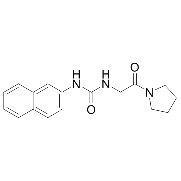 function and their role in disease etiology highlight the importance of developing phosphatase agonists and inhibitors. Unfortunately, phosphatases have historically been perceived as “undruggable” for several important reasons. First, phosphatases often control multiple signaling pathways and thus, inhibition of a single enzyme may not yield a specific cellular effect. Second, signaling cascades are generally controlled by multiple phosphatases and accordingly, blocking the activity of one may not sufficiently induce the desired modulation to a signaling pathway. Finally, and most importantly, phosphatase active sites display high conservation which hinders the ability to develop catalysis-directed inhibitors with any degree of selectivity. Despite these pitfalls, the emerging role of PTPs in human disease etiology has Screening Libraries necessitated a solution. Largely through use of structure-based drug design, several PTPs now represent promising targets for disease treatment. Most notably, bidentate inhibitors of PTP1B, implicated in type II diabetes and obesity, have been developed which span both the catalytic pocket and a second substrate binding pocket discovered.