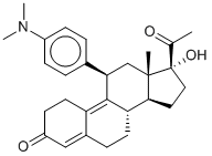
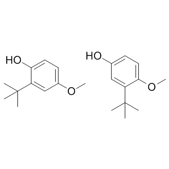
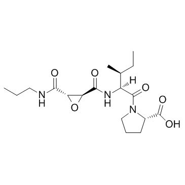
Since our understanding of ecosystem functioning depends on the data within a food chain, outdated, biased, or incomplete assessments of diet weaken our ability to predict how and why changes to ecosystems occur. Conventional methods of constructing food chains rely on diet studies which visually identify prey during feeding events or within stomachs, pellets, or feces. These techniques often suffer from MLN4924 Metabolic Enzyme/Protease inhibitor misidentification of similar-looking prey, underrepresentation of soft-bodied or small prey, and low taxonomic resolution, where prey cannot be precisely identified due to distance or digestion. Biochemical methods such as fatty acids, stable isotopes, and DNA have gained popularity in diet studies and can be used as a form of quality control for conventional techniques. However, these methods offer insight on diet in the longer term and are not necessarily well suited for prey species identification, particularly when there is little a priori knowledge of diet. The use of DNA within feces or stomach contents provides a snapshot of Tasocitinib predator diet and has not only been shown to provide a better estimate of diet than conventional methods, but can also provide species-level prey identification through the use of publicly available reference sequences. DNA-based techniques are advantageous in that they can be used either for comparative purposes for previously known diets or for de novo diet description. The application of DNA barcoding in diet studies has increased considerably with the advent of next-generation sequencing technology. It is now possible to identify even the rarest prey from multiple predators to species, genus, or family level in a single sequencing run while maintaining the ability to trace back each prey to the sample from which it came. NGS has been used in barcoding diet studies for fur seals, little penguins, slow worms, bats, leopard cats and tapirs. However prey identification from DNA in diet samples can be greatly influenced by technical issues including the uncertainty about the taxonomic diversity expected in the sample and the poor quality of the genomic DNA, particularly when extracted from fecal samples. Most DNA-based diet studies design multiple group-specific primers that amplify  the various prey types in predator diet, but these studies may fail to describe the full taxonomic range of the prey consumed. Universal primers can amplify and resolve species across a broad variety of taxa, making them a good, cost- and time-effective alternative to group-specific primers. Using multiple markers may also provide a broader taxonomic resolution of diet as different markers are not suitable barcodes for all taxonomic groups. In addition, PCR amplification of degraded DNA is more reliable when target fragments are small. Moreover, up to 90% of the sequences obtained from NGS can be less-degraded host DNA. The inclusion of primers to block host DNA amplification can increase the number of prey sequences significantly. Finally, it is also possible that the results may be misleading if primers can amplify the prey within the prey on which predators feed. This may skew the interpretation of how those species interact with the rest of the ecosystem. Thus a comparison of the diet of both predator and prey is warranted to assess the potential for detection of secondary consumption by DNA based methods. To date, no diet studies employing NGS have investigated multiple components of a food chain. Here we use Atlantic puffins of Machias Seal Island and their main prey, Atlantic herring as a model system to investigate how molecular methods can be used to describe food chains as these two species represent a simple food web where two conventional methods of studying diet have been employed. Puffin chick diet is known from hundreds of hours of observing adults provisioning their chicks as part of a long-term seabird research program.
Month: July 2019
Local microenvironment inhibited the differentiation of TAMs into the M1 phenotype and promoted their polarization into the M2 phenotype
Ohri et al. indicated that a higher M1/M2 ratio led to an increased 5-year survival rate in non-small cell lung cancer patients, but they also found that the altered M1/M2 ratio were resulted from the significantly increased infiltration of M1 TAMs and was therefore not the result of an imbalanced TAM polarization process. Later, Edin et al. indicated that it was the number of infiltrating M1 TAMs, rather than the change in M1/M2 ratio, that substantially influenced the prognosis of colorectal cancer patients. However, a study conducted by Zhang et al. found the opposite. The authors observed that an increased M1/M2 TAM ratio alone was sufficient to predict a better prognosis for lung cancer patients. Soon thereafter, Algars et al. corroborated the finding that it was the M1/M2 ratio itself, rather than altered densities of the infiltrating M1 or M2 TAMs, that predicted a reduced long-term incidence of cancer relapse or hepatic metastasis in patients with colorectal cancer. Therefore, our results validated the findings of Zhang et al. and Algars et al. We demonstrated that an altered  M1/M2 ratio alone is an independent prognostic indicator for ovarian cancer patients, which also implies that the chemotactic effect on monocytes/macrophages is not necessary for some immunosuppressive factors, such as MUC2, to influence patient prognosis. Moreover, considering that the reduced M1/M2 ratio has the highest HR, this pathological factor might have played a major role in the adverse outcome of MUC2++/+++ ovarian cancer cases. It was previously reported that cancer-derived MUC2 could initiate intracellular signaling by binding to the macrophage scavenger receptor on the surface of infiltrating monocytes/macrophages, promoting the upregulation of intracellular COX-2 gene expression in these cells. In this study, we re-examined this hypothesis in 102 cancer specimens. We found that most of the TAMs, which were closely surrounded by MUC2+ cancer cells, exhibited upregulated COX-2 expression. Moreover, we noted that this phenomenon was particularly significant in the high MUC2 expression group. These findings indicate that the local MUC2 expression level can help to determine the intratumoral density of COX-2+ TAMs, which implies its clinical significance. It has been known that COX-2 overexpression is associated with the increased synthesis of PGE2 in macrophages. The release of PGE2, which is a proinflammatory factor, can induce increases in the expression of VEGF, MMP, multi-drug resistance 1 and B-cell lymphoma 2 in the surrounding cancer cells, resulting in an improved vascularization of the cancer tissue and a reduced rate of apoptosis as well as an enhanced rate of metastasis and drug resistance in the cancer cell population. Hence, the existence of PGE2-releasing TAMs is generally an unfavorable prognostic factor. Because PGE2 is easily Foretinib degraded in the immunohistochemical labeling MK-2206 2HCl Akt inhibitor process, we had to immunohistochemically examined the COX-2 expression status in TAMs instead and used the obtained data for completing a Kaplan-Meier analysis. The result indicated that the COX-2 overexpression of TAMs was indeed associated with poor prognosis in the enrolled patients, suggesting that MUC2 also impaired patient survival via altering the local density of COX-2+ TAMs. In a seminal study, Torroella-Kouri et al. demonstrated that PGE2 could downregulate the transcriptional activity of NF-��B in immature monocytes and macrophages, which in turn reduces the expression levels of key M1-phenotype genes, such as iNOS and IL12. Recently, Nakanishi et al. confirmed that the administration of celecoxib, a selective COX-2 inhibitor, induced M2-polarized TAMs to become M1-polarized TAMs.
To validate the hypothesis that extra-cellular miRNAs confirmed the absence of cellular fraction
In addition to their association with microvesicles/exosomes and non-vesiclar molecules componants, very recent report showed that miRNAs may present in blood plasma in association of High Density Lipoprotein and delivered them to recipient cells with functional capabilities. In the present study, in addition to the exosomal fraction, we demonstrated that miRNAs were also coupled with non-exosomal fraction of the follicular fluid. Subsequently, we have confirmed the presence of miRNAs in both exosomal and nonexosomal fraction of the follicular fluid using the Human miRNome PCR array platform. Despite using a heterologous approach, the quantitative real time PCR analysis shows detection of the majority of miRNAs in both fractions of follicular fluid indicating the cross species conservation feature of miRNAs between human and bovine as it has been observed in wide range of species. Of the total of 750 miRNAs in the PCR array panel, a total of 509 and 356 miRNAs were detected in exosomal and non-exosomal fraction of follicular fluid respectively. Among the detected miRNAs, 331 were commonly found in both fractions, while 178 and 25 miRNAs were detected only in the exosomal and non-exosomal fraction of follicular fluid, respectively. This shows exosome mediated transport of miRNAs is the dominant pathway in bovine follicular fluid compared to the non-exosomal way as observed previously in blood plasma and saliva. To explore the possible association of exosomal and non-exosomal miRNA expression with oocyte growth in the follicle, we examined the relative miRNA expression level in exosomal and non-exosomal fractions of bovine follicular fluid collected from follicles containing growing and fully grown oocyte. Subsequently we found 25 and 32 miRNAs to be differentially regulated in exosomal and non-exosomal fraction, respectively between two oocyte groups. The higher number of up-regulated miRNAs in both exosomal and non-exosomal fraction of the follicular fluid of growing oocyte group may indicate the presence of a higher degree of Ibrutinib transcriptional activity during the growth phase of the oocyte. In order to evaluate the potential role of these differentially expressed miRNAs, the target genes were predicted bioinformatically and their biological function and gene ontology was determined. The most dominant categories enriched by predicted genes are related to transcription and transport which may  indicate that, growing oocytes have higher degree of transcription and translation resulting in efficient RNA management and storage in oocytes as maternal resource. Furthermore, the most significant pathways enriched by predicted targets for up-regulated exosomal miRNAs include ubiquitinmediated pathway, neurotrophin signaling, MAPK signaling and insulin signaling pathways. All these pathways are known to be involved in ovarian follicular growth and many CX-4945 developmental processes. The ubiquitin mediated pathway is known to modulate oocyte meiotic maturation, early mitotic division in developing embryos and plays important role in many cellular processes. While neurotrophin signaling pathway is reported to be important in regulation of oogenesis and follicle formation, the MAPK signaling mediates LH-induced oocyte maturation and its activation in cumulus cells is appears to require the permissive effect of the oocyte itself. Similarly, the overrepresented pathways in non-exosomal fraction were WNT signaling pathway and pathways in cancer and focal adhesion. WNT molecules are glycoproteins involved in fetal ovarian development and adult ovarian function including follicular growth, oocyte growth or maturation, steroidogenesis, ovulation and luteinization. Moreover, the higher number of common pathways in both exosomal and non-exosomal fraction may indicate that, miRNAs associated with either exosomes or Ago2 has a complementary function in follicular microenvironment.
Development is essential for coordination of folliculogenesis and triggering of different signaling molecules
Members of TGFB, insulin and WNT signaling family, growth factors such as GDF9 and BMP15, and hormonal regulation of FSH, LH that are also crucial for oocyte growth and developmental competence. During in vitro maturation and fertilization, a fully grown oocyte has better competency than a growing oocyte. Oocyte developmental competence is defined as the ability of an oocyte to resume meiosis, 3,4,5-Trimethoxyphenylacetic acid cleave following fertilization, develop to the blastocyst stage, induce a pregnancy and bring offspring to term with good health. The enzyme glucose-6-phosphate dehydrogenase is minimally active in the fully grown oocytes and present at higher level in growing oocytes. The enzyme G6PD can convert the Brilliant Cresyl Blue stain from blue to colorless; thus, growing oocytes will have a colorless cytoplasm while the fully grown ones remained blue. With that BCB staining of COC could be used as a method of screening oocytes for their growth status in many species including cattle and sheep. The development of COC to competent status is taking place in follicular microenvironment in which various signal transductions and molecular interactions are taking place between the surrounding cells mediated by the follicular fluid. Follicular fluid is a product of both the transfer of blood plasma constituents that cross the ��blood-follicle barrier�� and of the secretory activity of granulosa and thecal cells. It has been recognized as a reservoir of biochemical factors useful as noninvasive predictors of oocyte quality. Follicular fluid provides an important microenvironment for oocyte maturation and contains hormones such as FSH, LH, GH, inhibin, activin, estrogens and 4-(Benzyloxy)phenol androgens, pro-apoptotic factors including TNF and Fas-ligand, proteins, peptides, amino acids, and nucleotides. Follicular fluid is at least partly responsible for subsequent embryo quality and development and has some important oocyte-related functions including maintenance of meiotic arrest, protection against proteolysis, extrusion during ovulation and as a buffer against adverse haematic influences. As follicular fluid is derived from plasma and secretions of granulosa and theca cells, it is likely that products within follicular fluid may play a role in follicle growth and oocyte developmental competence. Exosomes have been postulated to play an important role in cell�Ccell communication, either by stimulating cells directly by surface expressed ligands or by transferring molecules between them. However, the mode of exosome-cell interaction and the intracellular trafficking pathway of exosomes in their recipient cells remain unclear. Exosomes are small membrane vesicles that are released into the extracellular milieu upon the fusion of multivesicular bodies with the plasma membrane. Unlike other  cell-secreted vesicles, exosomes are more homogenous with a size range from 40-100 nm in diameter. Exosomes contain a characteristic composition of proteins, and express cell recognition molecules on their surface that facilitates their selective targeting of and uptake by recipient cells. They are natural carriers of variety of coding and non-coding RNA, including microRNAs, which can be transported over large distances through blood to recipient cells and induce de novo transcriptional and translational changes in the target cells. These findings support the idea that exosomes might constitute an exquisite mechanism for local and systemic intercellular transfer not only of proteins but also of genetic information in the form of RNA. Currently, the role of exosomes, present in bovine follicular fluid, in transporting extra-cellular miRNAs within follicular environment and their contribution to follicular growth and oocyte maturation are unknown. During the dynamic phase of follicular development and oocyte maturation, miRNAs play an important role by coordinating.
Both exosomal and non-exosomal protein preparations were negative for CYC which is exclusively expressed
In a spatial and temporal specific manner. Several studies found that Albaspidin-AA miRNAs are not only present in cells but also in different body fluids including plasma, serum, urine, saliva, milk and semen, and those miRNAs are commonly termed as extra-cellular miRNAs or circulating miRNAs. Extra-cellular miRNAs are found to be remarkably stable in plasma despite high RNase activity in extracellular environment, suggesting that circulating miRNAs may be protected and bypass the harsh conditions in extracellular environment. The dominant model for extra-cellular miRNA transport and stability is associated with exosomes in different biofluids. In addition a significant portion of extra-cellular miRNAs are also associated with non-exosomal structures including Argonaute 2, the effector component of the miRNA-induced silencing complex, that mediates mRNA repression in cells. The discovery of miRNAs in body fluids, such as serum and plasma, and their remarkable stability opens up the possibility of using them as noninvasive biomarkers of disease and therapy response. So far, the presence of these extra-cellular miRNAs and their potential role in follicular microenvironment is not known. The present study was conducted to investigate: 1) the presence of extra-cellular miRNAs in exosomal and non-exosomal fraction of follicular fluid; and 2) differential expression of extra-cellular miRNAs in follicular fluid derived from growing and fully grown oocyte to postulate their role in oocyte growth. This study revealed the presence of extra-cellular miRNAs in bovine follicular fluid and found that the majority of them are associated with exosomes. Moreover, extracellular miRNAs are also present in non-exosomal part of follicular fluid. By comparing growing vs. fully grown oocytes, we found that both exosomal and non-exosomal fractions of follicular fluid carry a distinct set of differentially regulated extra-cellular miRNAs. Furthermore, exosomes can be taken up by surrounding follicular cells and subsequently increase the level of the endogenous miRNA in follicular cells. The supernatant of the samples were collected without disturbing the exosome pellets, and exosome pellets were resuspended in 200 ml of DPBS. Both the exosomes and exosome-depleted supernatant were further used for total RNA including small RNA fractions and protein Mepiroxol isolation to be used for miRNA PCR array analysis and detection of marker proteins, respectively. Recently miRNAs have been detected in extracellular environment mainly in different bio-fluids and their spectra could reflect altered physiological and pathological conditions. To our knowledge, this study is the first report on the presence of extra-cellular miRNAs in bovine follicular fluid, which may differ in its composition depending on the growth status of the oocytes. The results of the present study clearly support our hypothesis that miRNAs are also present in bovine follicular fluid being associated with either exosomes or non-exosomal structures and their signature may represent the altered physiological conditions in the follicular microenvironment. Various studies have demonstrated that circulating miRNAs are coupled  with either exosomes or AGO2 protein complex are able to bypass the high RNases activity in blood stream. By using a systemic approach, we have revealed that there are at least two populations of extra-cellular miRNAs in follicular fluid, namely exosomes and non-exosome associated, in which the former represent the majority of circulating miRNAs in follicular fluid. The specificity of exosome isolation was confirmed by the presence of CD63 in isolated exosomes. To support the hypothesis that along with exosomes AGO2 protein complex may also carry a sub set of miRNA in follicular fluid, we checked the presence of AGO2 protein complex in the non-exosomal fraction of follicular fluid.