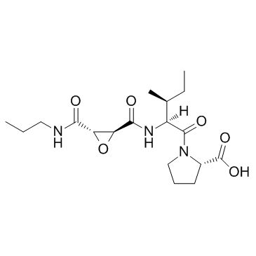
In a spatial and temporal specific manner. Several studies found that Albaspidin-AA miRNAs are not only present in cells but also in different body fluids including plasma, serum, urine, saliva, milk and semen, and those miRNAs are commonly termed as extra-cellular miRNAs or circulating miRNAs. Extra-cellular miRNAs are found to be remarkably stable in plasma despite high RNase activity in extracellular environment, suggesting that circulating miRNAs may be protected and bypass the harsh conditions in extracellular environment. The dominant model for extra-cellular miRNA transport and stability is associated with exosomes in different biofluids. In addition a significant portion of extra-cellular miRNAs are also associated with non-exosomal structures including Argonaute 2, the effector component of the miRNA-induced silencing complex, that mediates mRNA repression in cells. The discovery of miRNAs in body fluids, such as serum and plasma, and their remarkable stability opens up the possibility of using them as noninvasive biomarkers of disease and therapy response. So far, the presence of these extra-cellular miRNAs and their potential role in follicular microenvironment is not known. The present study was conducted to investigate: 1) the presence of extra-cellular miRNAs in exosomal and non-exosomal fraction of follicular fluid; and 2) differential expression of extra-cellular miRNAs in follicular fluid derived from growing and fully grown oocyte to postulate their role in oocyte growth. This study revealed the presence of extra-cellular miRNAs in bovine follicular fluid and found that the majority of them are associated with exosomes. Moreover, extracellular miRNAs are also present in non-exosomal part of follicular fluid. By comparing growing vs. fully grown oocytes, we found that both exosomal and non-exosomal fractions of follicular fluid carry a distinct set of differentially regulated extra-cellular miRNAs. Furthermore, exosomes can be taken up by surrounding follicular cells and subsequently increase the level of the endogenous miRNA in follicular cells. The supernatant of the samples were collected without disturbing the exosome pellets, and exosome pellets were resuspended in 200 ml of DPBS. Both the exosomes and exosome-depleted supernatant were further used for total RNA including small RNA fractions and protein Mepiroxol isolation to be used for miRNA PCR array analysis and detection of marker proteins, respectively. Recently miRNAs have been detected in extracellular environment mainly in different bio-fluids and their spectra could reflect altered physiological and pathological conditions. To our knowledge, this study is the first report on the presence of extra-cellular miRNAs in bovine follicular fluid, which may differ in its composition depending on the growth status of the oocytes. The results of the present study clearly support our hypothesis that miRNAs are also present in bovine follicular fluid being associated with either exosomes or non-exosomal structures and their signature may represent the altered physiological conditions in the follicular microenvironment. Various studies have demonstrated that circulating miRNAs are coupled  with either exosomes or AGO2 protein complex are able to bypass the high RNases activity in blood stream. By using a systemic approach, we have revealed that there are at least two populations of extra-cellular miRNAs in follicular fluid, namely exosomes and non-exosome associated, in which the former represent the majority of circulating miRNAs in follicular fluid. The specificity of exosome isolation was confirmed by the presence of CD63 in isolated exosomes. To support the hypothesis that along with exosomes AGO2 protein complex may also carry a sub set of miRNA in follicular fluid, we checked the presence of AGO2 protein complex in the non-exosomal fraction of follicular fluid.