To these surfactants than polarized epithelial cells. We interpret this result to mean that TX-100, DDPS and SDS act mainly at the level of the plasma membrane of the cells probably by causing structural changes at the level of the membrane or even its dissolution, as expected at concentrations close to the surfactant CMC. On the other hand, the cationic surfactants are probably toxic at a more subtle level, toxicity being at concentrations that are not sufficient to cause significant damage to the physical integrity of the membranes. These effects could even be at the intracellular level, conditioned by membrane partitioning and/or translocation across the membranes. The highly ordered apical membranes of fully polarized cells are the only membrane exposed to the surfactants in confluent polarized cell cultures and intact epithelia whereas non-polarized cells have significant amounts of less-ordered membrane domains exposed. For the homologous series of cationic surfactants examined, the results show that the toxicity to mammalian cells was not linearly dependent upon the surfactant hydrophobic chain length. This observation may have complex reasons related to different affinities of the surfactant for the different, possibly multiple, sites of their action. Without precise information concerning these affinities, something that we are working to obtain, any further discussion of this aspect would be speculative. The effect of the polar head group of the cationic surfactants was also evaluated; C12BZK and C12PB, which have the larger polar head groups and more delocalized charge, were between 2 to 5 times more toxic than C12TAB in all cell lines tested. Delocalized charge on the surfactant head group makes its ionic radius considerably larger and reduces the work required for translocation of the polar group from one side of the membrane to the other. Though there have been a very large number of reports concerning the disinfectant properties of surfactants, their mechanism of action is still not fully understood. Attempts to use surfactants in STI prophylaxis Catharanthine sulfate relied upon their capacity to destroy viral 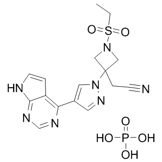 and bacterial membranes but did not seem to take into Benzethonium Chloride account, what in hindsight appears all too obvious, that if they destroyed those membranes they would also destroy the membranes of cells of the vaginal epithelium. However, destruction of cell membranes is not the only mechanism of surfactant toxicity as is evidenced in the case of cationic surfactants. As argued above, their toxic effects probably do not involve gross disassembly of the cell membrane but rather some more subtle effects. Candidate mechanisms that have been proposed in the literature include modulation of membrane curvature elastic stress and consequent reduction of membranebound protein activity, alteration of the electrostatic surface potential of membranes, or interaction with anionic polymers in the cytoplasm or cell nucleus following translocation across the cell plasma membrane. Cationic surfactants are known to bind strongly to DNA and RNA and induce drastic conformational changes in the structure of these polymers. The cell viability results obtained with the MTT assay correlated well with the observed release of LDH from the cells. However, in the case of C10TAB and C12TAB, the LD50 values obtained with the LDH leakage assay were slightly but significantly higher than with the MTT assay. This can be explained by the nature of each assay: the LDH leakage assay, which evaluates the loss of intracellular LDH and its release into the culture medium, is an indicator of irreversible cell death either due to cell membrane damage directly caused by the surfactants or due to loss of plasma membrane integrity posterior to cell death due to reasons that have nothing to do with direct membrane damage by the surfactants. On the other hand, the MTT assay evaluates the metabolic capacity of the cell in reducing MTT to formazan. Our results suggest that the molecular targets of the CnTAB surfactants may be different depending on the length of their hydrophobic chain.
Month: June 2019
Cmyc is the only gene of the four original iPS reprogramming factors that is represented in a stemness on module
To test for coordinate regulation of gene homologs or modules across different stem cell types, we developed a pattern recognition algorithm capable of combining the results of any number of experiments to identify significantly and recurrently up- or down-regulated genes and gene modules in stem cells. To obtain a global overview of stem cell expression patterns, we assembled a compendium of data from 30 different studies assaying gene expression of 49 stem cell populations representing twelve different types of stem cells including hematopoietic, retinal, neural, embryonic, and intestinal. From each of the 49 datasets we collected genes up-regulated in stem cells into a stem cell gene list and, where available, a corresponding set of genes up-regulated in differentiated cells into a differentiated gene list. The use of gene lists facilitated the straightforward integration of results from the variety of experimental test platforms, which has proven effective for meta-analysis compared to alternative approaches. A permissive, or poised, chromatin structure may underlie stem cell multipotency. S-MAP detected several chromatin-related modules Tulathromycin B associated with stemness including those involved in imprinting, chromatin-dependent silencing, heterochromatin and the nuclear lamina, which may indicate widespread suppression of lineage-associated genes. Stemness contributors include the Chd/Smarca family, nucleosome assembly protein like proteins; and histone variants H2afz and H2afv. Indeed, Chd1 was recently shown to be important in ESC multipotency by regulating chromatin structure; other Amikacin hydrate family members may fill this function in adult stem cells. S-MAP also revealed a number of Wnt signaling modules with alternating patterns of specificity in different stem cell types. Wnt-related stemness modules include the secreted Frizzled-like proteins of the Sfrp family; the Frizzled receptors; a subfamily of the TCF/LEF transcriptional regulators; the Enhancer of Split/Groucho-related Tle factors; and both alpha- and delta-catenins. Whether Wnt signaling regulates stem cell maintenance or differentiation has been extensively debated, and some genes have both inhibitory and activating abilities dependent on the state of Wnt signals. While functional consequences cannot be resolved by expression data alone, our analysis demonstrates that all stem cells tightly regulate the Wnt pathway at the transcriptional level. The identified S-MAP patterns provide specific candidates for functional interrogation in different stem cell types. DNA repair is uniquely critical in stem cells as mutations accumulated in stem cells amplify in differentiated daughters. SMAP identified many stemness modules associated with DNA repair, including the Terf, p53, and Rad families. Terf1 and 2 are highly stem cell-specific and are expressed in alternating patterns in different stem cells. S-MAP classified the p53 family as a stemness-on module. While p53 was found to be expressed in several tissues, p63 was upregulated in gastric and intestinal cells consistent with its known role in the development and maintenance of epithelial stem cells. Likewise, Brca1 and its homolog Mcph1, the Msh family of proteins, and a Rad/Dmc module were detected as stemness modules by S-MAP. Several transcriptional regulators, including the Myb family of oncogenes, were among the highest scoring stemness-on modules. c-myb was enriched in hematopoietic stem cells, consistent with its known role in differentiation control, as well as neural, embryonic,  intestinal and retinal stem cells. a-myb complements the expression of its partner genes by significant upregulation in gastric stem cells, while b-myb was up-regulated in liver and trophoblast stem cells. The Pbx and Id families have also been implicated in maintaining stem cell function. The myc family displayed a strongly complementary stemness expression pattern. c-myc has been implicated in reprogramming of differentiated cells into pluripotent cells.
It also implies that functional synapses more dynamic in younger preparations and become more stable
We used relatively young cultured neurons after 2�C3 weeks in vitro, when there is still a fairly high level of spontaneous synapse formation. Thus our results may be relevant to activity-dependent mechanisms of synapse formation during late-stage development as well as  synaptic plasticity. Although many studies have shown that synaptic components are dynamic, it has not been known whether they assemble and disassemble at random or fixed sites, and whether plasticity alters the number or dynamics of those sites. We have found that synaptic components assemble at fixed sites that are much more stable than the components themselves. In addition, quantitative modeling suggested that rapid and long-lasting changes in the number of those sites can account for most of the changes in puncta and structures during potentiation. By contrast, our data could not be fit by changes in the rates of assembly or disassembly of the puncta or structures in the model. That result differs from several previous studies that reported a change in the rate of turnover of synaptic components during functional plasticity. Ginsenoside-F2 However, a change in the number of sites could look like a change in the rate of turnover, and might at least partially account for those results. Fixed sites where puncta and structures assemble and disassemble have not previously been described, perhaps because most studies follow puncta or structures over a single cycle of assembly and disassembly. However, the sites are similar to “pause sites” on axons where synaptic protein transport Ginsenoside-F5 vesicles pause and where presynaptic terminals and stable contacts with postsynaptic filopodia preferentially form. Some of the puncta in our study could be transport packets of synaptic proteins, but two types of evidence suggest that most of our sites are not pause sites. First, we found that the appearance and disappearance of puncta of synaptic proteins is largely due to aggregation and disaggregation, rather than pausing. Second, whereas the pause sites do not require contact with a postsynaptic neuron, we found that puncta of pre- and postsynaptic proteins generally assemble and disassemble at shared sites. That result implies that the sites are located at points of contact between neurons, and must involve some sort of local communication between the neurons.
Changes in the small structures during potentiation there are increases in spine
Clusters or puncta of both postsynaptic glutamate receptors and presynaptic vesicle-associated proteins, as well as sites where the pre- and postsynaptic puncta colocalize. Within tens of minutes there is outgrowth of new pre- and postsynaptic structures, and by 0.5 to 15 hrs new synapses are formed. Homatropine Bromide Moreover, the early presynaptic alterations appear to depend on retrograde signaling from the postsynaptic cells, as occurs during the early stages of de novo synapse formation. These results suggest that the structural alterations during early-phase Catharanthine sulfate potentiation may act as tags or initial steps in a program that can lead to stable synaptic growth during late-phase potentiation. To begin to explore these ideas, we have examined changes in synaptic puncta and structures during long-lasting potentiation in hippocampal neurons. Early-phase potentiation is accompanied by rapid increases in puncta of presynaptic and postsynaptic proteins, which are due to aggregation of protein from a more diffuse background and are dependent on NMDA receptor activation and actin polymerization but not on protein synthesis. To investigate the possible contribution of these events  to late-phase potentiation, we have now addressed three questions: Do these early changes persist and become protein synthesis dependent? What is the nature of the changes? How might they contribute to the late phase? Our results suggest that the onset of potentiation is accompanied by rapid and long-lasting increases in the number of sites where synaptic puncta and structures are assembled. These new sites are much more stable than the puncta or structures themselves, and may contribute to the transition between the early and late phase mechanisms of plasticity by serving as seeds or tags for the protein synthesis-dependent assembly and maintenance of synaptic components during the late phase. To attempt to address that question, we divided the colocalized structures into ones that overlapped and ones that were adjacent to synapsin-IR puncta. Under control conditions, both overlapping and adjacent structures continually assembled and disassembled. However, almost all of the changes in structures during potentiation were attributable to changes in the overlapping structures. There was a gradual protein synthesis-dependent increase in adjacent structures, but no difference between the glutamate and control groups.
While these findings conciliate the results from data integration verification with those from gene signature evaluation
Each set had clinical follow-up information in form of censored time to event data, the event being either “overall survival” or “relapse-free survival” or both. The goal was to extract a gene signature from a training set that can be used to predict disease outcome for patients in the testing set. The gene signature we used consisted of a set of genes plus corresponding Cox coefficients derived by univariate fitting of the expression values to the survival data on the training set. Gene signatures were built from the individual or merged data sets. The accuracy and robustness of prediction were evaluated by 10-fold cross-validation. Reproducibility as defined above was analyzed by training a signature from one or several complete data sets and testing its performance on complete independent validation sets. Data sets were merged in their original numerical representation using two different data integration methods: ComBat and Z-score normalization. Two signature performance measures were computed in each experiment: time dependent Receiver-Operator Characteristic Area Under the Curve and the hazard ratio of the Echinatin predicted  risk scores relative to the survival data in the testing set. Note that the latter required stratification of the testing set patients in a high and low risk groups. In total, we analyzed 1324 breast cancer samples from public data sets generated with three microarray technologies. To the best of our knowledge, this study is the largest one evaluating the potential benefits of data merging in a quantitative OS/RFS patients risk prediction framework. They also reveal the limited usefulness of the data intermingling test, which in this case provides a misleading Pimozide picture of the variance retained after data integration. Noting that the gene signatures built from subsets of GSE4335 or Vijver showed higher prediction accuracies in cross-validation than the gene signature built from the merged data set, we investigated how the performance could possibly be improved by selective data integration. To assess the reproducibility of the gene signatures’performance derived from the merged data sets, the prediction accuracy was evaluated in a leave-one-data set-out manner. In each step, one complete source data set was set aside as testing set while the predictor was built from the merged remaining sets.