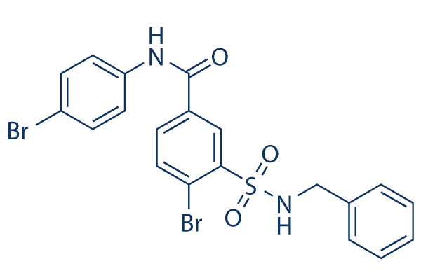
In rats, removal of the visual cortex during the first two postnatal weeks results in the rapid, massive death of Butenafine hydrochloride neurons in the dLGN with complete loss of the nucleus by adulthood. TUNEL- and activated caspase-3-positive cells were seen in the dLGN 24 to 72 hours following a lesion of the visual cortex, but were rarely observed 7 days after the lesion.  These results indicate that caspase-3 activity accompanying axotomy-induced cell death in the dLGN of young rats and mice is initiated rapidly after Folinic acid calcium salt pentahydrate axotomy and peaks in intensity shortly thereafter. In contrast, in axotomized dLGN projection neurons in the adult rat fractin-IR was detected first in dendrites, the most vulnerable compartment of these neurons at 36 hours survival. Only after the intensity of fractin-IR had increased in both dendrites and cell somas at 72 hours survival was fractin-IR detected in cell nuclei, which often appeared condensed. It is perhaps not a coincidence that the dendrites of axotomized dLGN projection neurons degenerate progressively during the first three days after axotomy. At 72 hours survival, approximately 60% of the dendrites of axotomized dLGN projection neurons have been lost, and cell somas have begun to atrophy and display nuclear condensation. These results indicate that the temporal progression of caspase-3 activity in axotomized dLGN projection neurons follows with a delay the same sequence of structural changes that are observed in these neurons; specifically, the structural changes seen in axotomized dLGN projection neurons and caspase-3 activity appear first in the dendrites of the injured neurons, and then in the next two days advance to involve the cell soma, preceding the death of the injured neuron. In an axotomized dLGN projection neuron, the progression of cytoskeletal degeneration and caspase-3 activity from dendrites to the cell soma is a roadmap leading to the death of the injured neuron. Interrupting this roadmap may offer an opportunity to protect axotomized neurons from dying. With this in mind, we investigated interrupting the roadmap with a single administration of FGF2 at the site of the cortical lesion to determine qualitatively and quantitatively if this affected the time course and extent of the dendritic degeneration in axotomized projection neurons in the rat dLGN. The present results clearly demonstrate that a single administration of FGF2 significantly reduces, but does not prevent, the degeneration of dendrites in these axotomized neurons. As mentioned, when the dendrites of injured dLGN projection neurons degenerate, the ability of these neurons to receive information from other cells and participate functionally in neuronal circuits is compromised. When axotomized projection neurons in the dLGN of the rat lose more than 50% of their dendrites, they frequently die. By contrast, when dLGN projection neurons retain more than 10 dendrites and a dendritic arbor with a cross-sectional area at least 20% of normal size, some projection neurons survive up to 7 days after axotomy. Thus, maintaining a minimum number of dendrites and a dendritic arbor of minimal dimensions appears to be associated with the survival of dLGN projection neurons after axotomy. Therefore, a reduction in dendritic degeneration following axotomy may play an important role in protecting injured dLGN projection neurons from dying. Administration of FGF2 in vivo has been shown to reduce neuronal death in the adult brain triggered by axotomy, excitotoxicity, MPTP treatment, and traumatic injury.